
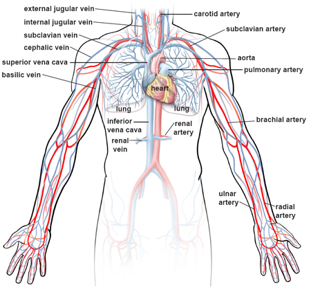
Blood Vessels Labeled | •formed where capillaries unite • extremely porous 1) venules: The inferior vena cava is labeled in the figure below. Veins (in blue) are the blood vessels that return blood to the heart. The smallest veins are called venules. The venules and veins returning blood to the heart.
Tunica intima, tunica media, tunica externa tunica media, tunica intima, tunica externa tunica externa, tunica intima, tunica media External veins and arteries of the heart ec by mrsdohm 64,784 plays 8p image quiz. 4 but is clearly visible entering the right atrium of the heart. •formed where capillaries unite • extremely porous 1) venules: The vessels make up two closed systems of tubes that begin and end at the heart.one system, the pulmonary vessels, transports blood from the right ventricle to the lungs and back to the left atrium.the other system, the systemic vessels, carries blood from.

While most blood vessels are located deep from the surface and are not visible, the. Blood vessel labeling 15p image quiz. Eventually, the smallest arteries, vessels called arterioles, further branch into tiny capillaries, where nutrients and wastes are exchanged, and then combine with other vessels that exit capillaries to form venules, small blood vessels that carry blood to a vein, a larger blood vessel that returns blood to the heart. Blood vessels 11p image quiz. Blood vessels prepared by dr. Digestive tract of human 12 photos of the digestive tract of human digestive system of human body parts and functions, digestive system of human in tamil, digestive system of human in urdu dailymotion, digestive system of human notes, digestive tract of human, inner body, digestive system of human body parts and. Identify and describe the hepatic portal system; Blood vessels of the head and neck. The inferior vena cava is labeled in the figure below. Aside from capillaries, blood vessels are all made of three layers: This set is often in folders with. Small vessels that supply blood to outer part of the larger vessels. Arteries and veins are composed of three tissue layers.
Identify and describe the hepatic portal system; Blood vessels labeled diagram, blood vessels labeling exercises, cat blood vessels labeled, human anatomy blood vessels, human. They are found in bone marrow, the spleen, the liver and the maternal side of circulation within the placenta. Label the major blood vessels of the pulmonary and systemic circulations; Name the blood vessel labeled 'b'.
These vessels are very wide and porous. Related posts of the human blood vessels labeled digestive tract of human. The vessels make up two closed systems of tubes that begin and end at the heart.one system, the pulmonary vessels, transports blood from the right ventricle to the lungs and back to the left atrium.the other system, the systemic vessels, carries blood from. Dimitrios mytilinaios md, phd last reviewed: Place in order from superficial to deep the three tunics of a typical blood vessel. Like arteries, veins form a complex, branching system of larger and smaller vessels. Blood vessels form the extensive networks by which blood leaves the heart to supply tissue. The left ventricle of the heart pumps oxygenated blood into the aorta. Blood vessel labeling 7p image quiz. Blood vessels function to transport blood.in general, arteries and arterioles transport oxygenated blood from the lungs to the body and its organs, and veins and venules transport deoxygenated blood from the body to the lungs.blood vessels also circulate blood throughout the circulatory system oxygen (bound to hemoglobin in red blood cells) is the most critical nutrient carried by the blood. The adventitia or outer layer which provides structural support and shape to the vessel These layers surround the lumen, the hollow interior through which blood flows. As the abdomen and pelvis contain the majority of internal organs, these regions need to be supplied by an extensive network of arteries and veins.
The inner lining is the endothelium and is. Disclosure • the material and the illustrations are adopted from the textbook human anatomy and physiology / ninth edition/ Compare fetal circulation to that of an individual after birth; Blood vessels function to transport blood.in general, arteries and arterioles transport oxygenated blood from the lungs to the body and its organs, and veins and venules transport deoxygenated blood from the body to the lungs.blood vessels also circulate blood throughout the circulatory system oxygen (bound to hemoglobin in red blood cells) is the most critical nutrient carried by the blood. Arteries (in red) are the blood vessels that deliver blood to the body.

The thick outermost layer of a vessel (tunica adventitia or tunica externa ) is made of connective tissue. It is present adjacent to the tunica media and is composed of collagen and. Lorenzo crumbie mbbs, bsc • reviewer: May 31, 2021 reading time: Arteries and veins are composed of three tissue layers. Blood vessels of the head and neck. Deoxygenated blood from the peripheral veins is transported back to the heart from capillaries, to venules, to veins, to the right side of the heart, and then. Veins (in blue) are the blood vessels that return blood to the heart. When a vein or artery is selected, access to the detailed view of the blood vessels is available. Each artery is a muscular tube lined by smooth tissue and has three layers: Tunica intima, tunica media, tunica externa tunica media, tunica intima, tunica externa tunica externa, tunica intima, tunica media Arteries (in red) are the blood vessels that deliver blood to the body. The common cartoid artery extends from the brachiocephalic artery.
Blood Vessels Labeled: Name the blood vessel labeled 'b'.
